 Written by: Dr. Tuba Çelebi Kayhan, Dermatologist
Published:
Short answer
Mole removal is taking a mole out under local anaesthetic because it looks suspicious, gets rubbed or is a cosmetic concern. After a dermoscopy check, surgical excision or shave removal is chosen to suit its structure, size and site, and the tissue goes for pathology. In Denizli, moles are removed at our clinic by Dr. Tuba Çelebi Kayhan.
At a glance
| Before deciding | Mole examination with a dermoscope |
|---|---|
| Methods | Surgical excision, shave removal |
| Numbing | Local anaesthetic |
| Removed tissue | Sent for pathology |
| Freezing and cautery | For benign lesions such as skin tags |
The decision to remove a mole rests on two questions: why should this mole come out, and how should it be removed? The first is usually answered by a mole check in Denizli; the answer to the second depends on the depth, size and position of the mole. The right method makes a pathology diagnosis possible and keeps the scar as small as it can be.
Which Moles Are Removed?
Most moles stay harmless for life and do not need to be removed. The situations in which we consider removal fall into three groups:
- Medical concern: a mole that shows an irregular structure under the dermoscope, grows, or changes colour or shape.
- Mechanical irritation: a mole that is constantly rubbed by a belt, bra strap, collar or razor, or that bleeds.
- Appearance: a mole on the face or another visible area that bothers you and has been found to be benign.
Not every raised spot people call a "mole" is a true mole. Skin tags on a stalk in areas of friction and the brown, rough seborrhoeic keratoses that come with age are different lesions, and they are removed in different ways.
Why Is an Examination Needed Before Mole Removal?
What the mole is determines the method. At the examination we look at the mole with a dermoscope and check for any suspicious structure. A mole that raises concern is removed completely, with a margin of healthy skin around it. For a mole that looks harmless and is being removed only for appearance, the method that keeps the scar smallest comes first.
The second benefit of the examination is identifying the moles that do not need removing. If you have come because one mole has changed, we also look at your other moles and discuss whether you need regular follow-up with dermoscopy.
What Are the Methods of Mole Removal?
Surgical excision (removal with stitches)
The mole is cut out through a spindle-shaped incision together with a thin margin of healthy skin, and the wound is stitched. Because all layers of the mole are removed, the pathologist can examine the whole mole. This is the preferred method for suspicious moles and moles that extend deep into the skin. It leaves a thin, line-shaped scar.
Shave removal
After a local anaesthetic, a mole that stands out from the skin is cut away level with the skin surface using a fine blade. No stitches are needed; the wound heals by forming a crust and usually leaves a flat, round mark. It is a suitable option for raised, benign-looking moles on the face.
Cautery and cryotherapy
Burning with heat and freezing with liquid nitrogen destroy a lesion but do not send any tissue for examination. For this reason they are used not on true moles but on lesions found to be benign at the examination, such as skin tags, seborrhoeic keratoses and warts. How they are used for these lesions is covered under cryotherapy and cautery treatments.
| Criterion | Surgical excision | Shave removal | Cautery / cryotherapy |
|---|---|---|---|
| Suitable for | Suspicious or deep moles | Raised, benign-looking moles | Skin tags, seborrhoeic keratoses, warts |
| Stitches | Needed | Usually not needed | Not needed |
| Pathology | The whole mole is examined | The removed part is examined | No tissue is sent |
| Resulting mark | A thin line | A flat, round mark | A small colour difference may remain |
Why Is a Removed Mole Sent for Pathology?
Dermoscopy gives a strong idea of a mole's structure, but the microscope gives the definitive diagnosis. Even with a harmless-looking mole, examining the removed tissue means the diagnosis does not rest on an estimate. For this reason, whenever we remove a mole we send the tissue for pathology, even if it was removed only for appearance. Preparing the sample and reviewing the result follow the same path as a skin biopsy.
Why Is Laser Mole Removal Not Suitable for Every Mole?
A laser destroys a mole by vaporising it or breaking up its pigment. No tissue is left to examine, so there is no way of finding out afterwards whether the mole was really harmless. For a suspicious mole, or one without a clear diagnosis, this is not an acceptable risk.
The second problem is depth. Mole cells can extend into the lower layers of the skin, and a treatment that works from the surface can leave these cells in place. When a mole reappears in the same spot, it can look irregular within the healing tissue and make dermoscopic assessment harder. This is why, for moles, we prefer methods that remove the tissue completely and allow it to be sent for pathology.
It is also possible to mistake a flat, light brown mark for a mole. If such a mark turns out to be a sun spot or freckle, it is a matter not for mole removal but for sun spot and freckle treatment, which uses different methods.
How Is a Mole Removed?
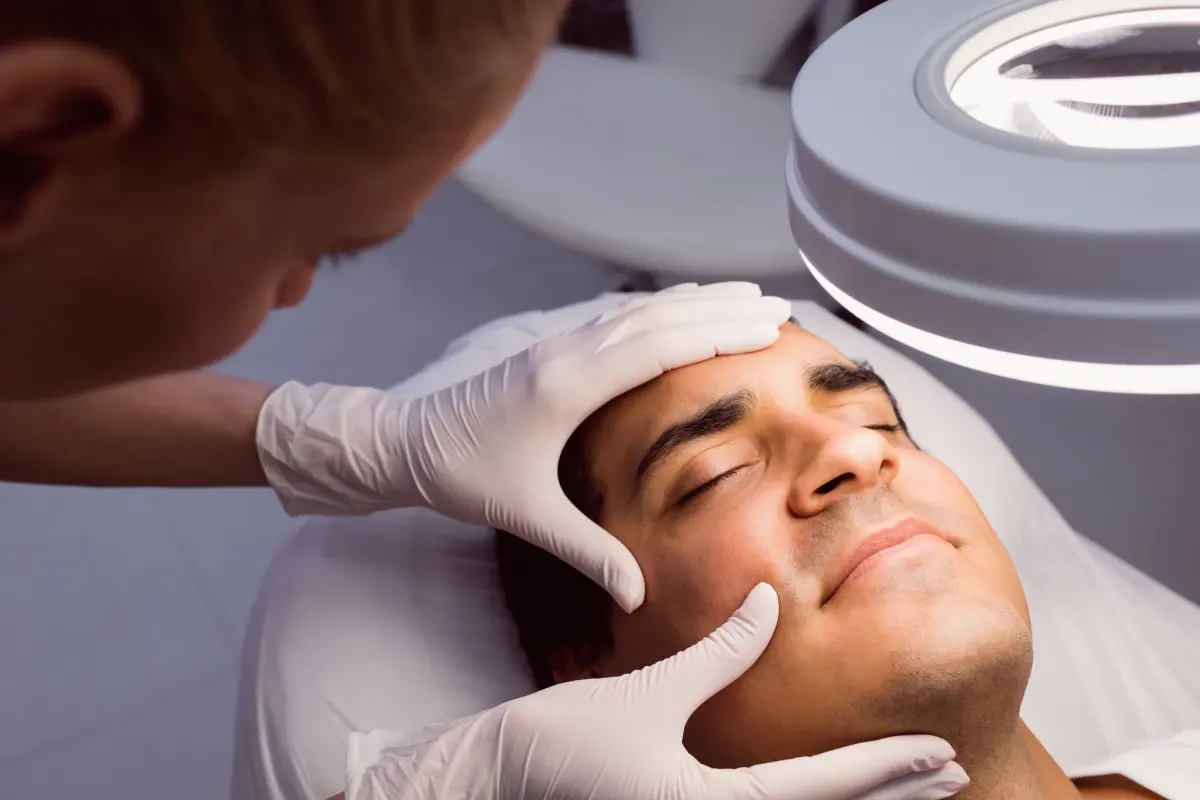
Before the procedure we ask about the medicines you take, especially blood thinners, and any known allergies. The site of the mole is marked, the area is cleaned and numbed with a fine needle. You feel a brief sting and burning during the injection; once the area is numb, you usually feel only touch and a slight pulling during the procedure.
The mole is removed with the chosen method, any bleeding is controlled, stitches are placed if needed and the area is covered. Afterwards you can go home or back to work on your own. Step by step, the removal follows the same course as excisional and shave biopsies.
Will Mole Removal Leave a Scar?
Any procedure that involves cutting or shaving the skin leaves a mark; the aim is to keep it small and discreet. The main factors that shape the scar are:
- Method: excision leaves a thin line, shave removal a flat, round mark.
- Size and depth of the mole: larger, deeper moles leave larger scars.
- Body area: on areas under tension such as the chest, shoulders and back, scars can widen and are more prone to raised healing.
- Personal tendency: if you have a history of keloid or raised scars, tell us before the procedure.
A new scar looks pink and noticeable in the first weeks, then fades and softens over months. Protecting it from the sun during this time is the most effective way to stop the scar from darkening.
What Should You Watch for After Mole Removal?
Wound care depends on the method and is explained to you at the end of the procedure. The general rules are: keep the dressing clean, do not pick the crust, avoid exercise that strains the area until any stitches are removed, and protect the healing skin from the sun. If you notice increasing redness, swelling, discharge or fever, call us.
We go through the pathology result together at the follow-up visit. If the result is benign and the mole has been removed completely, no further procedure is usually needed. If the result calls for further treatment, we discuss the plan in detail at the same visit.
Mole Removal in Denizli: How We Assess Your Mole
At our clinic, mole removal always begins with an examination: we look at the structure of the mole with a dermoscope and discuss with you whether it needs removing and which method suits you. We explain the likelihood of scarring and the healing process clearly before the procedure. For a mole examination at our clinic in Denizli, book an appointment.
Frequently asked questions
Can I have a mole on my face removed just for cosmetic reasons?
Can a removed mole come back in the same place?
If I have a hairy mole removed, will the hairs go too?
Is there a right season for mole removal?
Can I have more than one mole removed on the same day?
Which doctor should I see in Denizli to have a mole removed?
Sources
- Moles: Diagnosis and treatment — American Academy of Dermatology
- Moles — NHS
- Excision of skin lesions — DermNet
- Melanocytic naevus — DermNet
This page is for general information only and does not replace an examination, diagnosis or treatment. Please consult your doctor for advice about your own situation.